21208665
Subpopulations of equine blood lymphocytes expressing regulatory T cell markers.
Robbin MG, Wagner B, Noronha LE, Antczak DF, de Mestre AM.
Vet Immunol Immunopathol. 2011 Mar 15;140(1-2):90-101
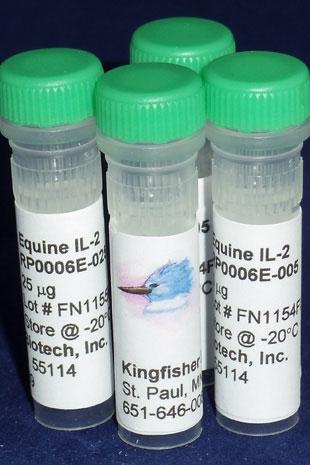
Applications: Cell Culture Stimulation
Abstract
Several distinct T lymphocyte subpopulations with immunoregulatory activity have been described in a number of mammalian species. This study performed a phenotypic analysis of cells expressing regulatory T cell (Treg) markers in the peripheral blood of a cohort of 18 horses aged 6 months to 23 years, using antibodies to both intracellular and cell surface markers, including Forkhead box P3 (FOXP3), CD4, CD8, CD25, interferon gamma (IFNγ) and interleukin 10 (IL-10). In peripheral blood, a mean of 2.2 ± 0.2% CD4+ and 0.5 ± 0.1% CD8+ lymphocytes expressed FOXP3. The mean percentage of CD4+FOXP3+ cells was found to be significantly decreased in horses 15 years and older (1.5%) as compared to horses 6 years and younger (2.7%), but did not differ between females and males and ponies and horses. Activation of peripheral blood mononuclear cells by pokeweed mitogen resulted in induction of CD25 and FOXP3 expression by CD4+ cells, with peak expression noted after 48 and 72 h in culture respectively. Activated CD4+FOXP3+ cells expressed IFNγ (35% of FOXP3+ cells) or IL-10 (9% FOXP3+ cells). Cell sorting was performed to determine FOXP3 expression by CD4(+)CD25(-), CD4(+)CD25(dim) and CD4(+)CD25(high) subpopulations. Immediately following sorting, the percentage of CD4+FOXP3+ cells was higher within the CD4(+)CD25(high) population (22.7-26.3%) compared with the CD4(+)CD25(dim) (17% cells) but was similar within the CD4(+)CD25(dim) and CD4(+)CD25(high) cells after resting in IL-2 (9-14%). Fewer than 2% of cells in the CD4(+)CD25(-) population expressed FOXP3. These results demonstrate heterogeneity in equine lymphocyte subsets that express molecules associated with regulatory T cells. CD4+FOXP3+ cells are likely to represent natural Tregs, with CD4+FOXP3+IL-10+ cells representing either activated natural Tregs or inducible Tregs, and CD4+FOXP3+IFNγ+ cells likely to represent activated Th1 cells.


