
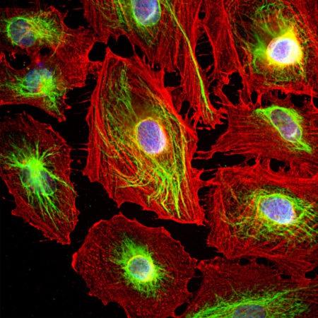
Figure-1: Immunofluorescence analysis of HeLa cells costained with rabbit pAb to vimentin,(34-1129), dilution 1:5,000, in green, and mouse mAb to Actin,(34-1002), dilution 1:500, in red. Blue is DAPI staining of nuclear DNA. The vimentin antibody stains the 10nm or intermediate filament network of the cytoskeleton. The antibody to actin labels the submembranous actin-rich cytoskeleton, stress fibers, and bundles of actin associated with cell adhesion sites.
