
Polyclonal Antibody to Peripherin
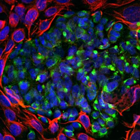
Figure-1: Immunofluorescent analysis of mixed fibroblast and PC12 pheochromocytoma cell culture stained with chicken pAb to peripherin,(34-1103), dilution 1:5,000 in green, and costained with rabbit pAb to vimentin,(34-1129), dilution 1:2,000, in red. The blue is DAPI staining of nuclear DNA. (34-1103) antibody reveals cytoplasmic filamentous staining in the small PC12 cells, while vimentin antibody stains intermediate filaments in the surrounding fibroblastic cells which do not express peripherin.
